
Leica LMD6 & LMD7
Laser Microdissection enables users to isolate specific single cells or entire areas of tissue.
Laser Capture Microdissection, Forensic Science, Pharmaceutical Chemical Engineering Research, RNA Analysis Workflow, DNA Analysis Workflow, Alzheimer Protein Research
Laser Microdissection Microscopes Leica LMD6 & LMD7
We move the laser, not the sample. And we use gravity for collection. That is why our LMD systems provide you with perfectly cut, contamination-free, analysis-ready dissectates.
Laser Microdissection (LMD, also known as Laser Capture Microdissection or LCM) enables users to isolate specific single cells or entire areas of tissue. Powered by a unique laser design and dynamic software, Leica LMD systems allow users to easily isolate Regions of Interest (ROI) from entire areas of tissue down to single cells or even subcellular structures such as chromosomes.
LMD is typically used in genomics (DNA), transcriptomics (mRNA, miRNA), proteomics, metabolomics, and even next generation sequencing (NGS). Researchers in neurology, cancer research, plant analysis, forensics or climate research rely on this method. Furthermore, LMD is a perfect tool for live cell culture (LCC), for cloning and re-cultivation, manipulation or downstream analysis.
We
Move the Laser, not the Sample
Try writing your name on a piece of paper by moving the paper instead of the pen. Hard to do? That’s why we move the laser, not the sample for laser microdissection.
Only Leica Microsystems uses high-precision optics to steer the laser beam along the desired cut line on the tissue with prisms. That means that the Leica LMD cuts perpendicularly to the tissue and leaves clean cuts of contamination-free isolates.
Precision.
Always.
You can cut with highest possible precision and speed
You can cut directly, real-time with "Move and Cut"
You get the best view for convenient movie documentation.
Gravity
for Cleanliness
Your downstream analysis relies on contaminate-free isolates. This is why Leica LMD systems collect dissectates via gravity. Their unique laser-guidance based dissection method preserves the integrity of your isolates – contact-free and contamination-free.
1, 2, 3 – contamination-free sample!
You choose the area of interest
You move the laser along the area to be cut out
The dissectate drops into the Petri dish and is ready for further analysis – 100% contamination-free
The real asset: Gravity always works.
Success
is Objective
You cut best with objectives that are dedicated and optimized to the task. Since the development and manufacture of optics have been among of our core competencies from the start in the 19th century, you can rely on the performance of our dedicated LMD objectives, our SmartCut series.
Choose
from a range of 10 dry objectives – from 5x to 150x
Use the unique 150x SmartCut objective to go into detail with high magnification and high resolution when required
Get a large field of view to cut large parts of samples in one piece with low-magnification objectives
Benefit from objectives with the highest possible transmission of laser light at 350 nm to cut tissue, bones, teeth, brain, plants, chromosomes and living cells – try them for your application!
The outstanding imaging performance our objectives provide goes without saying.
Same
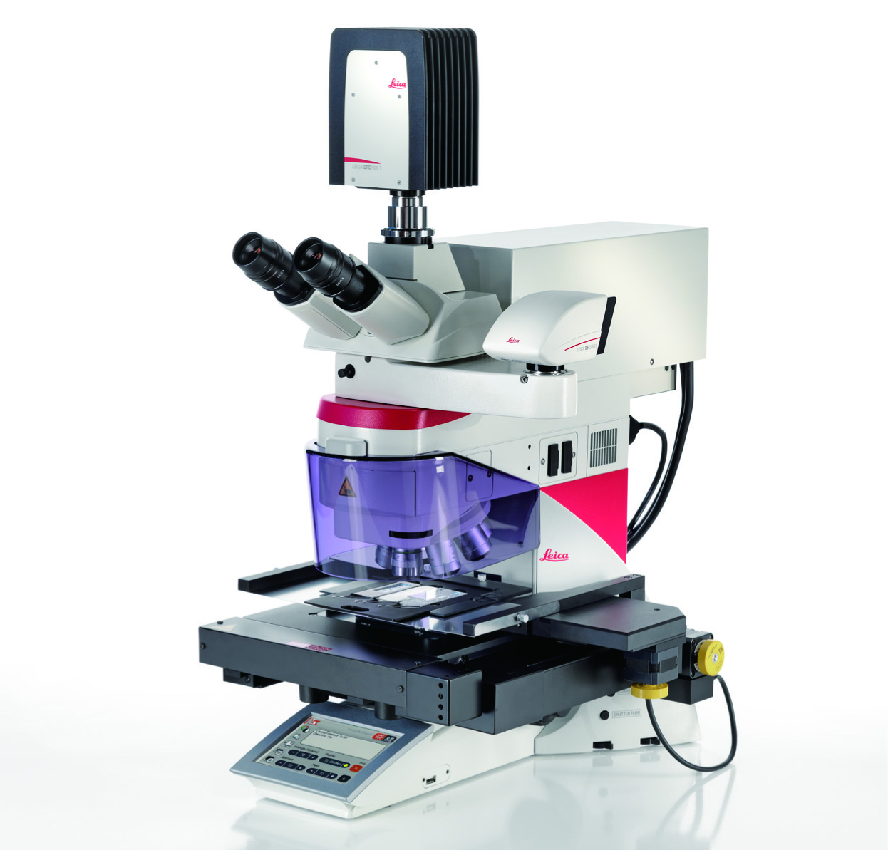
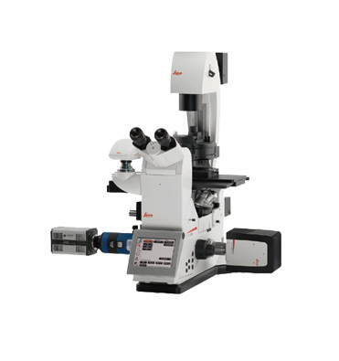
Principle, Two Systems
Choose your power: The difference between the Leica LMD6 and Leica LMD7 is the laser. It makes the Leica LMD6 the perfect tool for standard applications dissecting soft tissues such as brain, liver, or kidney. And it makes the Leica LMD7 ideally suited to dissecting any kind of tissue independent of its size or shape. It provides for greater flexibility with higher laser power and more laser controls than in the smaller system.
Two
systems for your discoveries
Leica LMD7 for highest expectations and most flexible use
Leica LMD6 for outstanding results in standard tissue dissection
Homogenous
Illumination
Light is crucial when you define your dissection area. This is why both the Leica LMD6 and Leica LMD7 are available with the traditional Halogen or LED illumination.
How
you benefit from LED illumination
You can see the well-lit sample in its natural colors, because LED illumination lights the sample homogenously and with a constant color temperature
You save time and money with LED illumination: LEDs require up to 90% less energy and have a lifetime of up to 25,000 hours – with this, instrument downtime due to bulb changes are a thing of the past
Do you prefer Halogen illumination? No problem!
If Halogen illumination for transmitted light is still your first choice, we can equip both systems with this. We provide it with internal constant color temperature control (CCIC) to avoid any image alterations from your traditional illumination technique, even if the system is used for laser microdissection independent applications.
Cutting-edge Laser Technology at a Glance
Leica LMD6 Leica LMD7
Wavelength 355 nm 349 nm
Pulse frequency 80 Hz 10-5000 Hz
Pulse length < 4 ns < 4 ns
Max. pulse energy 70 μJ 120 μJ
How the Leica LMD7 makes you more flexible
It integrates high energy per pulse and high and adjustable repetition rates in one system
You gain full control of the repetition to adapt the laser speed to a specific sample
You can use high-energy per pulse for thick and hard specimens
You benefit from high repetition rates for narrow cuttings and high speed
You control all laser parameters including laser aperture to achieve an optimal cutting line.
Save on Consumables!
Since the Leica LMD systems collect dissectates simply with gravity, you can use standard devices for their collection to all common molecular biology reaction devices such as 0.2 or 0.5 ml tube caps – which you already have in your lab. Collection devices can be dry or filled with reaction buffer or culture media for the LMD application.
Use membrane slides for optimal results with the "Move and Cut" mode: Cut your dissectate directly live. This method is called laser microdissection and is the most efficient and time-saving way to obtain your dissectate in best quality
Use the "Draw and Scan" mode to cut from normal glass slides, coverslips or DIRECTOR slides: This method is called laser ablation or dot scan dissection, which allows membrane-free collection.
Easy
Live Cell Cutting
If you work with live cells, you are used to working with inverted microscopes. Live cell cutting works smoothly with our LMD systems, even though the Leica LMD systems are based on an upright microscope.
You can dissect living cells in culture in order to re-cultivate, clone, or analyze single cells, colonies, or cell clusters
You can even attach a climate chamber to the LMD system
You can grow cells on petri dishes with PEN membrane or multi-well ibidi slides
You can collect the dissectate of living cell cultures into Petri dishes with or without PEN membrane, ibidi slides, or 8-stripe tubes for re-cultivation, or into collection devices such as PCR tube caps for analysis
Software
Makes LMD Easier
You are interested in your results, not in how to attain them? Then you will like the dedicated, workflow-oriented and intuitive application software. Easy to use and powerful, enabling you to select, dissect, and visualize the dissectates.
Get a specimen overview for better orientation
Guide the laser beam with mouse or touchscreen
Control laser and microscope
Record a time-lapse video
Add-on packages such as a database, automated cell recognition (AVC, pattern recognition), and several more at your service. Speak to your local Leica representative to find out what we have to offer!
Ultimately: Save time and effort
Slide
Solutions for Every Need
If you work in proteomics or metabolomics, the plasticizer or softener of membrane slides may interfere with your analysis. This is why Leica Microsystems offers different slide options for laser microdissection.
Use
any kind of membrane slides for genomics and transcriptomics
Use PET slides for certain applications in proteomics and metabolomics. PET is almost free of softeners.
Choose DIRECTOR slides for proteomics and metabolomics to work completely membrane free
High-Throughput
Capability
You need more throughput? The LMD systems can be configured with different kinds of stages. In addition to the standard devices you can choose the LMT350 ultra scanning stage, giving you fast, silent and ultraprecise navigation.
Moreover it is capable of holding collector vessels starting from 0.2 ml tube caps, through 8 and 12 cap-strips up to standard 96-well plates.
This enables you to do high-throughput experiments. Since you don’t need any special material but use regular 96-well plates, you can load them directly into standard PCR machines.